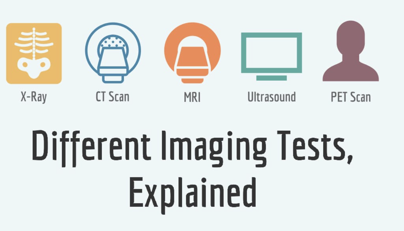
Understanding Imaging Tests for Joint Pain: X-rays, MRI, and What They Tell Us in Buffalo, NY 🔍
When persistent joint pain takes hold, one of the most common next steps in the diagnostic journey is an imaging test. For many residents of Buffalo, NY, facing this prospect, the process can feel confusing or intimidating. What’s the difference between an X-ray and an MRI? What are doctors looking for? And how do these images truly help in understanding what's causing their discomfort? Understanding the purpose of common imaging tests empowers you to be a more informed participant in your healthcare journey, providing clarity on how professionals in Western New York get to the heart of your joint pain.
Why Do Doctors Order Imaging Tests? The Diagnostic Puzzle 🧩
Before any imaging test is ordered, a thorough physical examination and a detailed review of your medical history are always the first and most critical steps. These foundational elements provide the clinical context—the "story" of your pain. Imaging tests then serve as powerful diagnostic tools, providing objective visual evidence to help confirm or rule out a suspected condition.
Imaging helps professionals:
- Visualize the Unseen: It allows doctors to look inside the joint without a surgical incision, assessing structures that cannot be seen or felt from the outside.
- Assess Severity: Imaging can help determine the extent of damage from conditions like arthritis, allowing for a more accurate understanding of the condition's stage.
- Differentiate Conditions: It helps distinguish between conditions that may have similar symptoms. For instance, is the knee pain from a bone fracture (seen on X-ray) or a meniscus tear (seen on MRI)?
- Plan a Path Forward: The information gained from imaging is crucial for developing a precise plan for managing your joint pain.
In Buffalo, where diverse joint issues are common, using the right imaging tool for the right situation is key to getting an accurate and timely understanding.
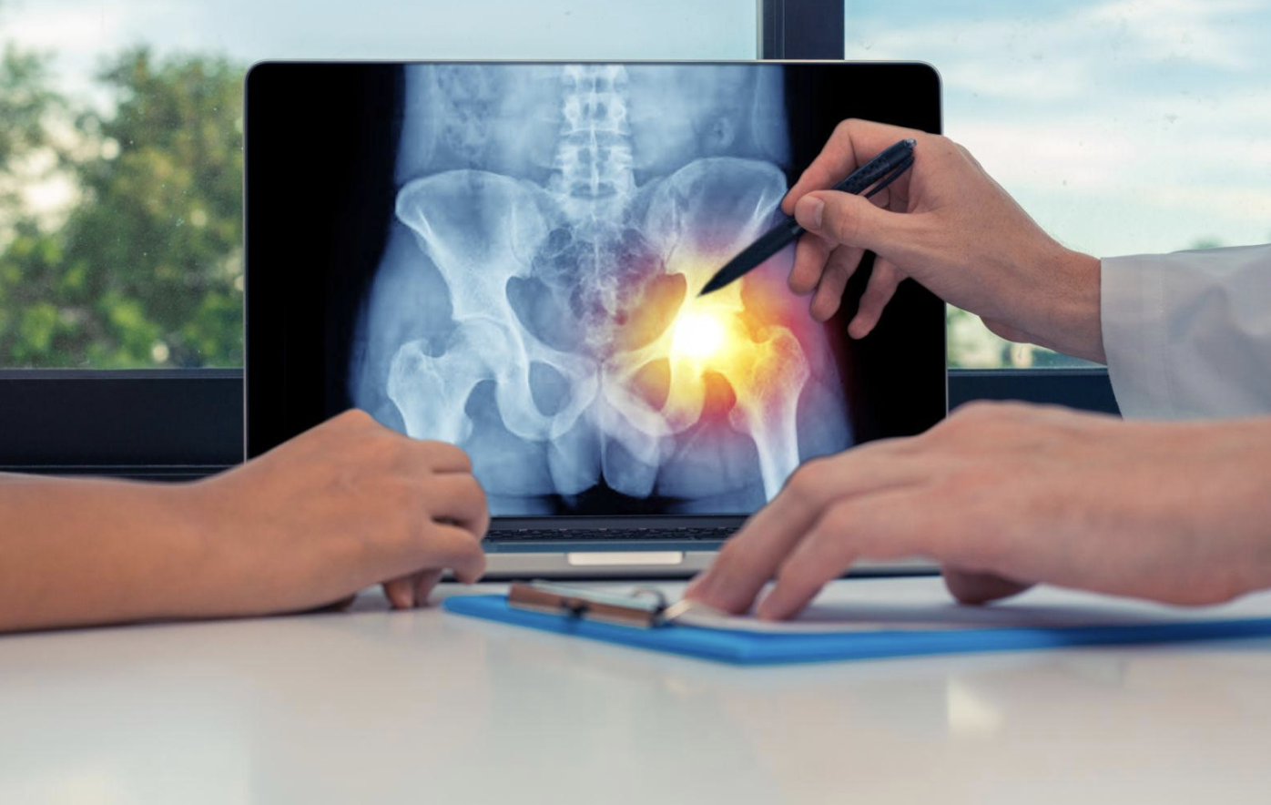
Imaging Test 1: The X-ray – Seeing the Bones 🦴
The X-ray is one of the oldest and most widely used forms of medical imaging. It's often the first test ordered for joint pain due to its accessibility and speed.
- What it is: An X-ray is a quick and painless test that uses a small amount of radiation to create images of the dense structures within your body. Because bones are very dense, they appear white on an X-ray, while soft tissues are not visible.
- What it shows:
- Bone Structure: X-rays are excellent for viewing bones. They can effectively diagnose fractures, dislocations, bone spurs (osteophytes), and other bone deformities.
- Joint Space: By showing the space between two bones, an X-ray can indicate cartilage loss. If the space between bones appears narrow, it is a key sign of conditions like osteoarthritis (OA).
- Bone Alignment: It can reveal issues with joint alignment, such as in conditions like a bunion or a severe deformity caused by arthritis.
- What it doesn't show well: This is a crucial point. X-rays are not designed to show soft tissues. Therefore, they cannot visualize cartilage directly, or tears in ligaments, tendons, or muscles.
- Why it's a first step: An X-ray serves as a great baseline to rule out common bone-related issues and to assess for advanced signs of arthritis. For a patient in Buffalo with persistent knee pain, an X-ray would be a standard first test to see if there is joint space narrowing or bone spurs.
Imaging Test 2: MRI – The Soft Tissue Detective 🔬
If an X-ray comes back normal but pain persists, or if a soft tissue injury is suspected, an MRI (Magnetic Resonance Imaging) is often the next step.
- What it is: An MRI is a non-invasive, painless test that uses a powerful magnetic field and radio waves to create incredibly detailed images of all the body's tissues. It does not use radiation. The process involves lying still inside a large machine that can be noisy (earplugs are provided!).
- What it shows:
- Soft Tissues: This is where MRI truly excels. It provides an unparalleled view of ligaments (like an ACL or PCL in the knee), tendons (like the rotator cuff in the shoulder or Achilles tendon in the ankle), cartilage, and muscles. This makes it the go-to test for diagnosing tears, inflammation (tendinitis, bursitis), or damage to these structures.
- Fluid and Inflammation: An MRI can clearly show fluid buildup within a joint (effusion) or inflammation in the soft tissues, which can be a key sign of an active condition.
- Spine and Disc Issues: MRI is the gold standard for visualizing conditions in the spine, such as herniated or bulging discs, nerve root compression, and spinal stenosis.
- Why it's ordered: An MRI is typically ordered when a soft tissue injury is suspected, such as a rotator cuff tear in a shoulder, a meniscus tear in a knee, or a disc issue causing nerve pain in the back.
- What it doesn't show well: While it shows bones, it is not as clear as an X-ray for certain bone-related issues like fractures. It is also not used to measure bone density.
Imaging Test 3: Ultrasound – The Live Look Inside 🔭
Ultrasound has emerged as a valuable tool for diagnosing and assessing joint-related issues, particularly soft tissue problems.
- What it is: An ultrasound is a non-invasive, radiation-free test that uses high-frequency sound waves to create real-time images. A hand-held probe is moved over the skin of the affected area, and images are displayed on a screen.
- What it shows:
- Soft Tissues in Motion: A key advantage of ultrasound is that it can provide dynamic, real-time images. A doctor can watch a tendon glide over a joint as the patient moves their arm, or see if a bursa is swelling.
- Inflammation: It is excellent for visualizing inflammation of tendons (tendinitis), bursae (bursitis), or fluid buildup.
- Cysts and Masses: It can help identify soft tissue cysts or masses.
- Guided Procedures: Ultrasound is often used to guide procedures, such as injections, ensuring a needle is placed with greater precision directly into the source of the problem.
- Why it's useful: Its quick, dynamic nature makes it an excellent tool for assessing specific, localized soft tissue issues. It's often used when an injury to a specific tendon or bursa is suspected.
Putting it All Together in Buffalo, NY 🧩
It’s crucial to remember that no single imaging test tells the whole story. An X-ray might show perfect bones, while an MRI reveals a significant soft tissue tear. Conversely, an MRI might show a lot of "wear and tear," but a patient's pain may be minimal. This is why the physical examination, the patient's symptoms, and the expert interpretation of the images are paramount.
The role of a specialist in Buffalo is not just to order tests, but to synthesize all of this information—your story, the physical exam findings, and the images—into a cohesive and accurate understanding. This holistic approach ensures that the right problem is identified and that the path forward is precisely tailored to your needs.
Seeking Expert Guidance for Joint Pain in Buffalo, NY 🧑⚕️
If you're living in Buffalo, NY, and experiencing persistent joint pain, seeking specialized guidance is the most important step you can take. While this article doesn't detail specific treatments, connecting with professionals who understand complex joint conditions and who know which diagnostic tools to use and how to interpret them is essential.
A comprehensive evaluation involves more than just ordering tests; it's about a partnership where you and your healthcare provider work together to piece together your health puzzle. For residents of Buffalo, NY, and throughout Western New York, gaining this precise understanding of your pain is the first step toward finding lasting relief.
Empower Your Health: Ask Questions About Your Images! 🌟
Imaging tests are powerful tools, but they are just one piece of the diagnostic puzzle. By understanding what each test is for and what it can and cannot show, you can be a more informed and empowered patient. Don't hesitate to ask your healthcare provider questions about your X-rays or MRI results; understanding your own body is the first step toward better health.